You go to your routine dental hygiene appointment, and the dental hygienist informs you that you need X-rays today. Even though the time can feel like flying, you may think, didn’t I just get these done? Or why do I really need them? And are they needed for dental cleanings?
X-rays (radiographs) are needed for dental hygiene appointments/teeth cleanings. X-rays are used to diagnose dental issues and also to study bone levels and health, teeth health, cancer, polyps, cysts, tumours, abscesses, impacted teeth, infections, missing teeth, and view developing teeth.
When I clean my patient’s teeth, I am constantly using multiple things to analyze my patient’s teeth. I use the clinical appearance of how the oral tissues look to my eyes, I use diagnostic tools such as gum probing measurements and X-rays, and I ask my patients questions to treat them accordingly.
Even though I use the patient’s x-rays at every appointment, that doesn’t mean the patient should have them taken at every appointment. There could be a couple of reasons why x-rays may need to be taken more often, and I go over all of those reasons below, so keep reading!
How I use dental x-rays for teeth cleanings
When I am scaling (cleaning) teeth, I often refer to the X-rays. I can see so much more than just with the naked eye. I use what I see on the X-ray to visualize how the teeth are shaped and create an image in my mind of what the tooth looks like underneath the gum line.
To be honest, cleaning teeth without x-rays can be like wearing a blindfold. I cannot visualize or see what is going on in the places I cannot see with my naked eye. When x-rays are up to date and recent, you get a better dental hygiene appointment and a lot more value out of it.
Periodontal disease
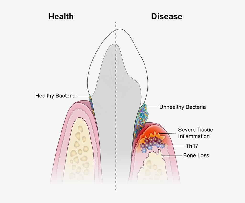
This is the BIGGEST reason that I need to use the x-rays for. Gum disease can be a little unpredictable, as sometimes it only affects a certain area/tooth or can be uneven around the mouth.
I can see where the bone levels are, between the teeth, and generally what type of bone loss is occurring.
Even though dental X-rays are so beneficial to treatment, sometimes dental offices take them too often and take too many. Below, I linked a post I wrote all about how often X-rays should and shouldn’t be taken and what you need to look out for!
Read Now: How Often Should You Get Xrays at the Dentist? DH Explains
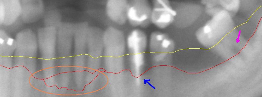
I use both the x-rays and a periodontal probe together to assess the bone levels around each tooth as dental x-rays are only 2D, giving us only some of the full picture.
And when a bone level could be quite low on a tooth, the respective gum tissue may be high, and to the naked eye, it looks like everything is healthy. But when looking at the x-rays and the gum measurements, a different story is painted. Also, the health of the bone is observed, and we can see if the top of the bone is in a more active state of destruction.
Gum disease can be increased by other things, not just poor oral health. A lot of people use a CPAP machine with sleep apnea that can contribute to dental disease. You will be surprised how one medical condition can have such a profound impact on oral health! Click the link below!
Read now: Dry Mouth With CPAP: Prevent and Eliminate Dental Problems
The angle of teeth/root shape
Everyone is unique, and even though our teeth follow a certain anatomical shape, everyone has differences.
While I am cleaning underneath the gum line, I use the x-rays to see the angle and shape of the tooth. I visualize this in my mind, so I can contour the instrument appropriately around each tooth. This makes the cleaning much more comfortable for the patient. Because I know the root shape, the instrument has less chance of injuring the soft tissue.
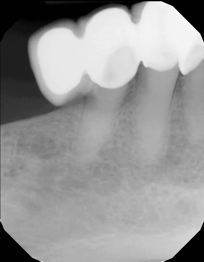
Calculus (tartar) formation
The hard deposits on the teeth form from the bacteria and plaque when it gets calcified to the teeth. It will never stop forming; for some people, it can form quite quickly.
The calculus forms both underneath the gum line, and above the gum line, on all surfaces of the teeth. The roots of the teeth have indents and small areas that are hard to reach with the floss and toothbrush and will build up more calculus.
If the deposit gets large enough, it can be seen on the x-ray. This points out an area to me that needs to be focused on by both myself as the dental hygienist and the patient, who may need to focus on that area while they are doing their home oral care routine.
Dental hygiene appointments should be very thorough, and the length of them can vary. The link below will explain in detail the aspects of a dental hygiene appointment and how long it should be. Are your dental hygiene appointments long enough?
Read now: Scaling and Polishing: How Long a Typical Dental Cleaning Is
I also use the x-ray as a teaching tool for the patient to visualize what is going on in their mouth and have a better understanding. I find this can be motivating for the patients.
Dental work examples: fillings, crowns, bridges, implants etc.
It is so important for us hygienists to have a full picture of what is happening below the gum line, between the teeth, and everywhere we cannot see.
Dental work comes in all shapes and sizes, and knowing the contour or the edge of the dental work, provides me with valuable information.
If I can see on the x-ray that some of the dental work is overhanging the tooth, and there is quite a large edge to it, I know not to go too aggressive with the cleaning in that area to not damage the dental work. I will also notify the dentist about this, and if it is negatively affecting the patient’s oral health, it may need to be fixed or redone.
What the dentist looks for in dental x-rays
Decay
Even though dental hygienists are not allowed to diagnose decay, we know what it looks like on an x-ray and have enough knowledge to know what we are looking at. If I take an x-ray and see an area suspect of decay, I can use that information to my advantage.
I can educate the patient on flossing and home care to stop the cavity and remineralize it if it is small enough. This is, of course, after the dentist has diagnosed it.
The dentist will diagnose the decay and explain to the patient what needs to be done.
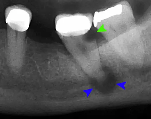
Disease
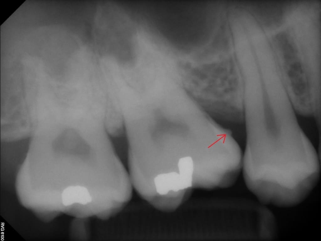
Apart from gum disease, evidence of other diseases is looked for on dental x-rays. These include abscesses and current infections.
In the picture under the decay subheading above, a dental abscess is indicated by the blue arrows.
It is extremely important to diagnose abscesses quickly because they can become very serious and could cross the blood-brain barrier and cause septic shock.
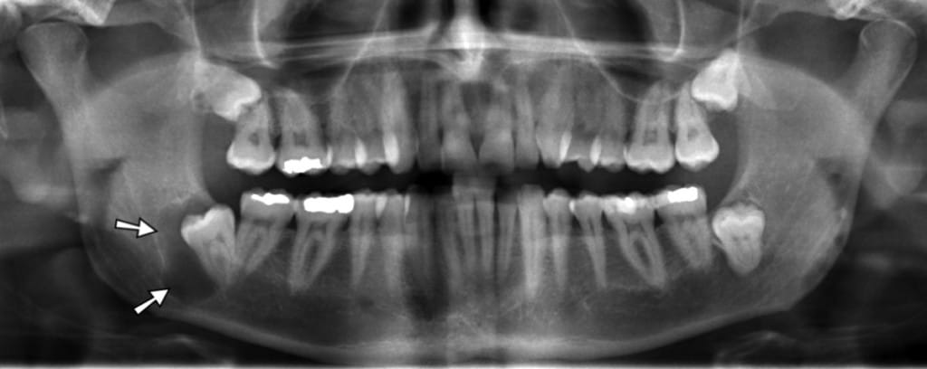
Nasal Polyps
Nasal polyps occur in the sinuses and can be seen on a panoramic x-ray. Usually, they are painless and caused by chronic inflammation from allergies, infections, asthma, and other conditions.
I once took a panoramic X-ray of a patient who was a 25-year-old male. We saw that he had quite a large nasal polyp, and I asked him if he ever had discomfort when flying.
He said yes, and he could not fly because the pain would be so bad. It really affected his quality of life, as travelling was limited by the pain he knew he would experience. Long story short, we recommended he go and see his family doctor and get a referral to see a specialist to have it treated. It gave the patient hope that he may be able to fly one day without being in immense pain in his sinus.
Cysts
Cysts can form pretty much anywhere in the head and neck, but we watch out for them in the jaw bone. They should be monitored regularly because they can seriously affect health.
Cancer
This is an important one. So many cancers can go undetected in the mouth for so long, and once they are noticed, it is often in the late stages of cancer and harder to treat, ending up with more severe repercussions.
Tumours
The effects on the hard tissues from tumours can be seen on dental x-rays. And if something is not looking quite right, we will refer to a specialist right away. It is important to catch these things early.
Abnormalities in tooth structure
Resorption
Teeth can resorb for multiple reasons. This is how baby teeth (with the help of the adult tooth underneath it) lose their roots, so only the crown is left when it falls out.
But in adult teeth, it can lead to premature tooth loss, and sometimes the cause is unknown. It most often occurs with braces, and the teeth are moved too quickly/aggressively. It can also occur will chronic inflammation of the nerve/pulpal part of the teeth.
Shape of tooth
Root shape is important if the tooth needs to be extracted or a root canal needs to be done. Roots that are misshaped, such as having curly roots, will be difficult to treat. Also, if the root/tooth is fractured, it can often be seen on an x-ray.
How often should I be getting dental x-rays?
There is a personal answer for everyone, and because some people have more dental issues, they may need more x-rays than someone who has not had a cavity in years and whose gum health is good. But you definitely do not need them every 6 months, or every year, unless there is a specific area that is being monitored, and if left can have a detrimental effect.
The following chart is a guideline only and should only be used as a tool, not a rule, when we take X-rays. Everyone should be assessed individually, and when there is a need to take them, where the benefits outweigh the risks, they should be taken.
| Situation | Frequency of dental x-rays |
| Child: Good oral health, no history of decay | Bitewings: Taken from the age of 6-7 (when adult 6-year molars erupt) Panorex: 6-7 years of age to check for all adult teeth |
| Child: Poor oral health/history of decay | Bitewings: Every 12-24 months, depending on the situation Panorex: 6 years of age |
| Teenager: No history of decay/good oral health | Bitewings: Every 24 months Panorex: Around the age of 15-16 to check on wisdom teeth |
| Teenager: A history of decay/poor oral health | Bitewings: Every 12-24 months depending on the situation Panorex: Around the age of 15-16 to check on wisdom teeth |
| Adult: No history of decay/good oral health | Bitewings: Every 24-36 months Panorex: Every 5-7 years |
| Adult: History of recent decay | Bitewings: Every 24 months Panorex: Every 5 years or as indicated |
| Adult: Specific area being watched | That specific area will be x-rayed in 6-12 months |
I know dental offices that take bitewings every year, regardless of patient or situation. And that is just wrong. For someone who has no history of oral disease or cavities and no other indicator to take X-rays, they should not be taken every year.
The reason why a lot of dental offices will take bitewings yearly is that insurance companies will cover the price, which means more money for the office. Putting money and greed over the patient’s health and safety is one of the worst things you could do.
I am so thankful I work for a dentist that cares about people more than the money that comes in.
Get a second opinion
If you think that you are getting too many X-rays, or something seems fishy, then get a second opinion. Truly, I really recommend second opinions. Not everyone knows everything, and also, I hate to say it because my passion is dental hygiene, but like anything else in life, money is sometimes put over people’s care.
Trust your gut and seek out advice from other professionals. If you tell your dental professional you want a second opinion, pay attention to their reaction. If it is something you need and is correct, they should encourage a second opinion.
How much radiation is from dental x-rays compared to other x-rays
The radiation dose measurement that we use is microsieverts and millisieverts. It really depends on the equipment that is used that determines how much radiation the patient is exposed to.
Older film-type x-rays use a lot more radiation to produce an image on the film vs the newer digital x-ryas that use an electronic sensor. The sensors are much more sensitive to the radiation.
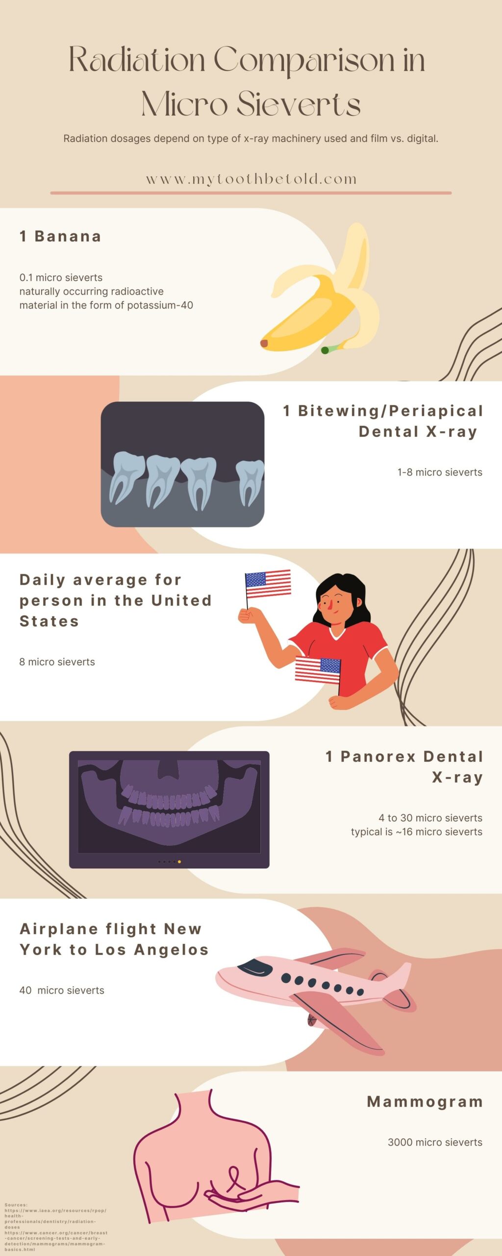
I love comparing the dosage of the x-rays that we take in the dental office to eating a banana. It really puts things into perspective how little radiation you are getting from dental x-rays.
However, you still don’t want to be taking them out of routine because you can. The ALARA principle still stands; however little the radiation is, it should always be minimized as much as possible.
Can X-rays be transferred to a new dental office?
X-rays can be transferred to a new office. Before switching to another office, find out when your last x-rays were taken, if possible. Or, you can get your new office to contact your old office to get them transferred. You will need to sign a release form permitting the X-rays to be transferred.
I am bringing this up because sometimes the new dental office will just go ahead and take new X-rays. But if you have recent X-rays from another office, you shouldn’t be having new ones taken.
Time, place, and a purpose
They are needed but not as often as you think- it’s not the same for everybody.
Dentistry has evolved much in recent years to a more patient-centred care approach. Not everybody is the same, and they should not all be treated the same.
The ALARA principle
The ALARA principle stands for “as low as reasonably achievable.” All X-rays cause some biological damage, even if a very small amount can come from dental X-rays.
Radiation is accumulative over a lifetime. And the radiation that you have been exposed to 10-20 years ago and your whole life stays with you. That is why dental x-rays should be limited and not taken on a routine basis.
Lead Aprons
Wearing a lead apron can protect the body from scatter radiation. Scatter radiation can have a different wavelength and have more of a risk of damaging the tissue. There are a lot of vulnerable tissues in the body that should be protected.
Even though dental x-rays are considered a very low amount of radiation, you still want to protect the body as much as you can, and go with the famous saying “better safe than sorry“.
Digital vs film-based X-rays

Many dental offices have switched to digital x-rays from film, and with good reason.
Digital x-rays offer a much lower amount of radiation than films and save time that could be used for educating and performing treatments on patients. It also completely takes away the need for dangerous chemicals. Old film-style X-rays needed dangerous chemicals to process, which are dangerous to the user and the environment.
Also, because the X-rays are digital and on the computer screen, they can be manipulated for better accuracy when diagnosing them.
I can show my patient a larger format where they have a problem, show them their bone levels and explain the dental treatment they may require.
Types of dental x-rays
Periapical
This type of x-ray is an image of the whole tooth, from the biting surface to the tip of the root (and a few mm past the root). These x-rays are often taken to see if there is an infection at the root tip and to check for root fractures. They can also be used to observe the sinus level and a few other things.
If there is evidence that makes the dentist want a periapical x-ray, we will take it. They are not done on a routine basis unless there is no access to a panoramic x-ray machine. However, a panoramic is a much better diagnostic tool than doing multiple periapical x-rays, and it is a lot less radiation, which is a bonus!
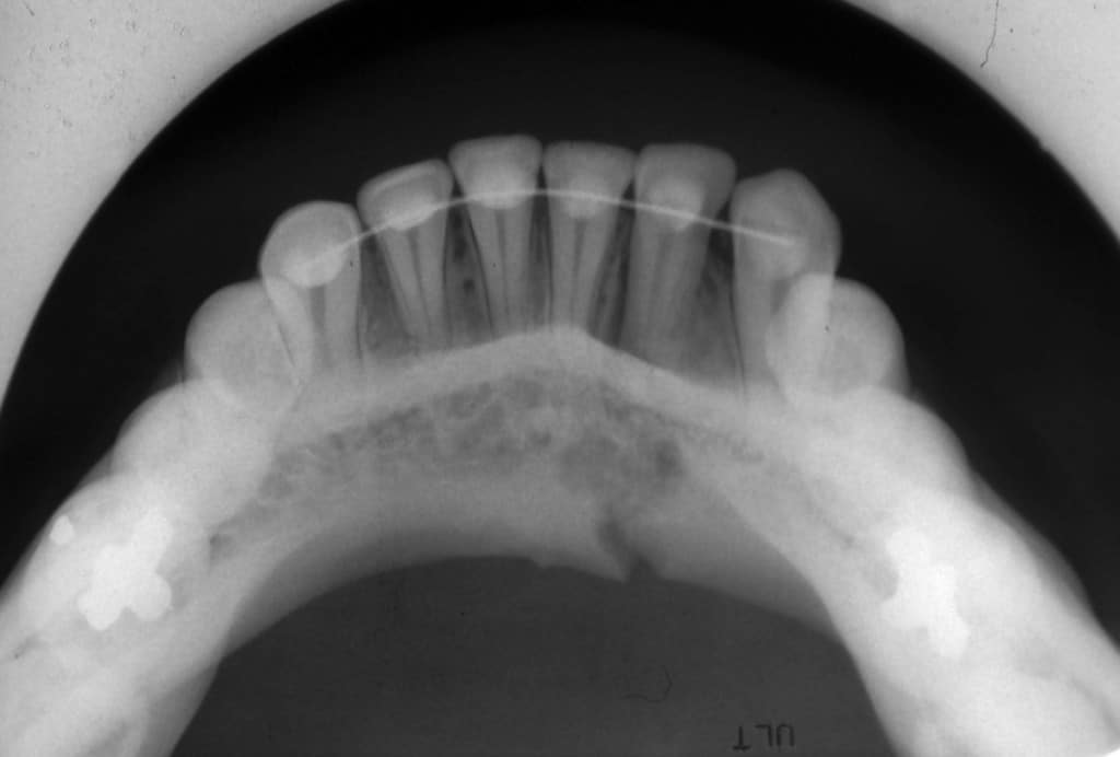
Bitewing (often referred to as cavity-detecting x-rays)
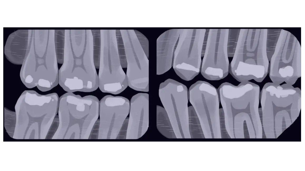
These x-rays are taken the most often, and their main purpose is to check for decay/cavities. The most common area where cavities form is right under between the teeth, where the two teeth touch together.
Again, it is important to have these because if decay is caught early, you can actually arrest it and stop it from progressing.
Panoramic (panorex)
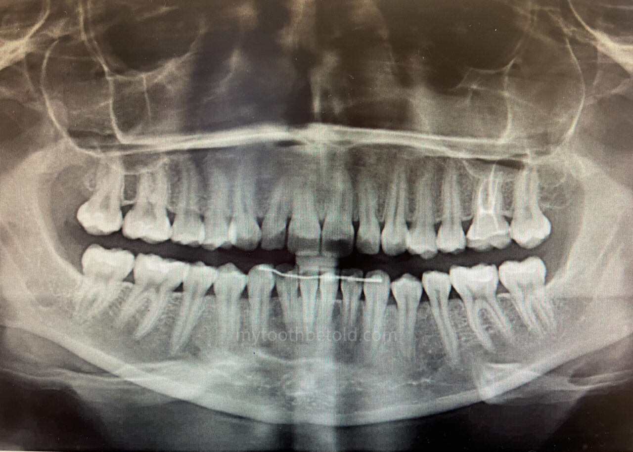
This x-ray is done more infrequently and is quite large. It takes a picture of both the top and bottom jaw, the nose and up to the eye sockets.
This x-ray should be done when a child is around 6-7 years old, specifically to check if all the adult teeth are present. There have been a couple of instances where we can see there is a missing adult tooth, and the parents are informed, and a plan can be developed.
It is important to check for all the adult teeth, so the appropriate plans can be made. How is the baby tooth going to be replaced, are braces probable, how long can the baby tooth be kept for?
As the patient gets older, around 14-16 years of age, we will take this x-ray again, and this time to check on the developing wisdom teeth (or if there are any missing), that way we can refer if we need to at the right time, for the wisdom teeth to be extracted.
Click the link below to find out why some people don’t have wisdom teeth. Is it evolution or another interesting reason?
Read Now: Why Do Some People Not Have Wisdom Teeth?
This x-ray is also taken to observe bone levels in those patients with periodontal disease. The insurance companies will need a copy of this x-ray to validate a person’s request for more dental cleanings.
Other dental X-rays
There are a couple of other types of x-rays that can be taken, but they are rarely used.
Occlusal X-rays
This type of x-ray is of one arch of the mouth and used to evaluate developing teeth in children but can be used in adults for certain diagnostic reasons.
Cephalometric x-rays
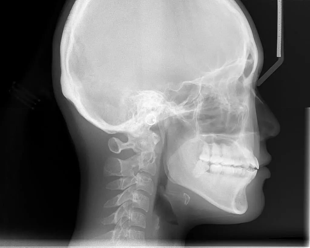
This type of x-ray is used by orthodontists to assess the side profile of the face. It is used when planning treatment and to see how the teeth come together on a side angle.
CBCT scan
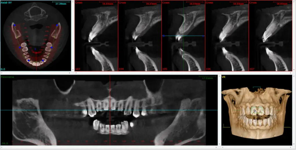
These are 3D images of the head and neck. Usually, they are used in more complex dental treatments such as implant therapy and oral surgery.
Click the link below to find out exactly how wisdom teeth are taken out. It’s a pretty cool and quick process!
Read now: How Are Wisdom Teeth Removed?
Sometimes if the wisdom teeth are quite close to a nerve, they will do a CT scan of the head and neck to know the exact location of the nerve and its proximity to the wisdom teeth. A 2D x-ray overlays a lot of structures, and it is hard to determine how close the nerve is to the tooth.
What type of radiation is used in dentistry
There are different types of radiation, and we use ionizing radiation in the dental setting. Ionizing radiation has more energy and directly affects the tissues of our bodies.
Radiation is all around us
This is just a recap of the information that is present in the charts and imaging in this post. Radiation is everywhere, and we are constantly exposed to it.
Some examples are our cell phones, living in stone/brick houses, and our means of travel. Even the elevation in which we live has an impact on how much radiation we absorb from space.
There is no escape. This thing that we all have to remember is; the dose is the poison, and do the benefits outweigh the risks?
Are dental X-rays harmful?
As I mentioned in the previous subheading, dental x-rays are ionizing radiation. Ionizing radiation can disrupt the tissue cells and the DNA in them. And all radiation is accumulated over time.
However, with dental x-rays, the wavelength that is sent out in a straight line is set to a specific spec and will pass through the tissue with extremely minimal damage. It is the scatter radiation that can cause more harm to the tissue. Because the wavelength is different, it affects the tissue cells more. This is why the patients wear a lead apron.
Are dental hygienists, assistants and dentists exposed to radiation?
Yes, we are. But, it is such a slight amount. The amount of radiation that is safe to be exposed to is way more than all the X-rays we even take. And we are not standing in the room with the patients.
The safe distance away from the source of the x-rays is six feet. So, that is why we always leave the room when we take the x-rays.
The walls and cupboards/shelving units are also lined with lead. Any scatter radiation that is produced gets blocked and absorbed by the lead.
Dental professionals can also wear dosimeters, which measure the amount of radiation that we are exposed to. They are organized by the health departments of the government and are sent in to be routinely checked.
I hope that this post has provided some insight to the dental world and x-rays.
Holly 🙂
Information sourced from;
https://pubmed.ncbi.nlm.nih.gov/18762634/
https://pubmed.ncbi.nlm.nih.gov/15897284/
https://www.ada.org/en/member-center/oral-health-topics/x-rays
https://www.nrc.gov/about-nrc/radiation/health-effects/info.html#dose
https://www.epa.gov/radiation/calculate-your-radiation-dose
https://www.epa.gov/radiation/radiation-sources-and-doses
